Hip and Elbow Dysplasia Screening

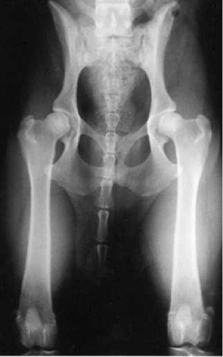
Figure 1: The difference between normal and dysplastic hips and the consequence of dysplastic hips leading to severe OA.
What is hip and elbow dysplasia (HD/ED)?
They are debilitating conditions of the hip and elbow joints whereby the joint structure doesn’t develop correctly (Figures 1 and 2). This leads to varying degrees of degenerative joint disease (DJD) which generally in older animals causes them to develop osteoarthritis (OA).
Unfortunately, we also see this in some younger animals; and are often causes of great expense to try to either surgically correct the deformity and/or extensive medical management of the pain and mobility impairment issues of the affected animals. In the worst cases, the animal’s pain is so extensive that they are unable to enjoy any significant quality of life and it leads to euthanasia.
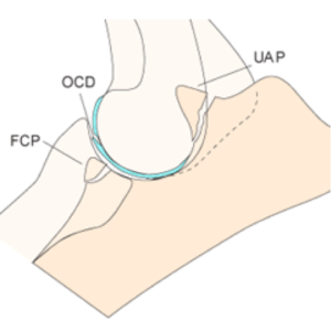
Figure 2: The three components of Elbow dysplasia are: Fragmented coronoid process (FCP); Osteochondrosis dessicans (OCD); and Ununited anconeal process (UAP).
It is widely accepted that HD/ED is not exclusively a genetic condition; and that especially in larger breeds, appropriate nutrition when growing and exercise management is also a critical component of the development of HD/ED.
Whilst it is critical to follow these guidelines in young large breed animals; these diseases also affect dogs of all sizes and breed and leads to DJD/OA changes at any age. It should be mentioned that in adult dog’s; appropriate nutritional, exercise and weight management is also a significant component of the development of DJD/OA.
Why screen for hip and elbow dysplasia?
The genetic component of HD/ED is very significant; and whilst nutrition & exercise management is very important it is critical that dog breeders aim to produce dogs with the best chance of having as little HD/ED within their lines as possible.
The Australian National Kennel Council (ANKC) has partnered with the Australian Veterinary Association (AVA) in a disease prevention program called Canine Hip and Elbow Dysplasia Scheme (CHEDS). This requires certain dog breeds to have had their hips and elbows screened for HD/ED before their litters can be registered. It is also strongly recommended to screen all animals for HD/ED as well as any other breed related genetic conditions that can be avoided by prudent breeding standards.
Doyalson Animal Hospital recommends screening all potential breeding animals for genetic conditions including HD/ED. We can assist in x-ray, DNA profiling and other techniques required to identify the conditions with the aim of reducing the incidence of these issues in breeding animals. Our expert veterinarians can also provide advice on interpretation of the results of these screening tests.
Which techniques are available?
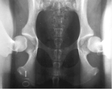
Whilst there are a number of techniques available for the assessment of dogs with HD/ED, the ANKC currently only recognise the system where a standard ventro-dorsal (VD) flexed hip and lateral flexed elbow x-rays are taken and assessed by a registered specialist radiologist (Figure 3). The AVA accredited radiologist will then allocate a score based on a number of set criteria and issue a report stating the individual numbers and total scores for both the hips and elbows. According to the breed there are acceptable score ranges but the general principal is to use breeding stock with the lowest scores and breed to try to reduce the incidence of HD/ED. These x-rays cannot be taken until the animal in question is 12 months old or older and female animals must not be actively cycling.
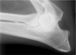
Figure 3: Standard VD extended pelvic and lateral flexed elbow x-rays.
PennHIP Screening Technique
In 1983 a new system for the prediction of HD was established and in 1993 the PennHIP organisation was born. They have developed a system for the evaluation of HD and prediction of OA development in dogs. This has been shown to be the most reliable screening system for HD and is backed up by many peer reviewed studies over past decades.
The PennHIP screening technique not only allows evaluation of DJD by utilizing the standard VD pelvic x-rays; but by using a specific set of compression/distraction views it can subjectively allow assessment of hip joint laxity which is the primary factor leading to DJD/OA in dogs (Figure 4).
The PennHIP system uses a research based set of measurements to establish a Distraction index (DI) to show the amount of hip laxity present. The lower the number the better with the aim to breed dogs with DI’s as close to 0.30 as possible.
Distraction View
Compression View
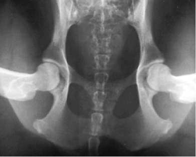
Hip-Extended View
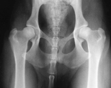
Figure 4: PennHIP screening technique showing the standard VD extended pelvic x-rays as well as the compression and distraction views. It the difference between the compression and distraction of the hips that allows for the Distraction Index to be calculated.
Why chose PennHIP screening?
PennHIP x-ray studies are only allowed to be performed by accredited veterinarians who have not only undertaken the course requirements but have had several PennHIP x-ray series evaluated to ensure the correct procedures are being undertaken. The x-rays are then sent to the PennHIP organisation in the USA where they are assessed by a specialist radiologist. All PennHIP x-rays taken around the world are sent to the same central location and this has allowed the establishment of a world-wide canine hip dysplasia database.
PennHIP xrays can be taken from 16 weeks of age. Animals can be screened at the same time as they are desexed if required. PennHIP hip x-ray assessment has been shown to be the most reliable predictor for hip laxity and HD.
How we can help
Dr Sally Smith is fully accredited to perform PennHIP as well as the standard hip and elbow dysplasia screening x-rays. We can perform these in house Monday to Friday. The process for either x-ray techniques requires the use of general anaesthesia hence a full day hospital stay is required. Animals can be dropped off between 8.30 and 9am and will be assessed by a veterinarian before admission into hospital; most animals are ready for collection from 4pm the same day.